Plate Nº 18 · recorded November 12, 2026
Neuroscience & MindReported finding
Meta-Analysis of 66 Studies Maps White Matter Differences in Autism
A meta-analysis of 66 studies covering 4,087 participants finds autistic individuals show altered white matter microstructure in nine of twelve major brain pathways.
By Marcus Bennett4 min read811 words
In brief
- The meta-analysis pooled 66 studies with 2,231 autistic and 1,856 typically developing participants, ages 2 to 50.
- Autistic individuals showed lower fractional anisotropy in 9 of 12 white matter tracts and higher mean diffusivity in 8.
- The largest effect appeared in the arcuate fasciculus, a language pathway, though the authors caution it may be inflated.
- Differences were smaller in studies with higher average IQ (seven tracts) and older samples (two tracts).
- The paper was published in Biological Psychiatry: Cognitive Neuroscience and Neuroimaging.
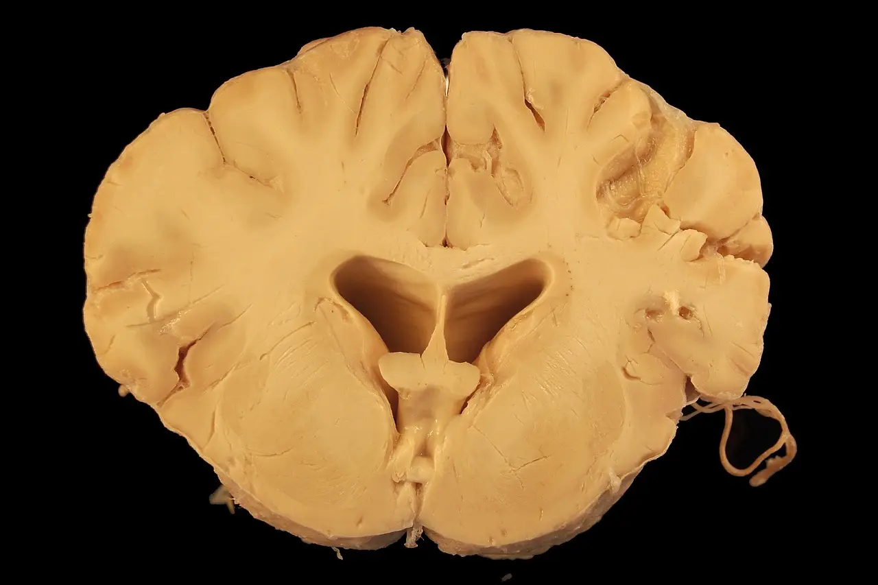
A meta-analysis of 66 brain imaging studies has found that autistic individuals show moderately altered white matter — the brain's long-range wiring — in most of the major pathways researchers examined. Compared with typically developing individuals, autistic participants showed lower fractional anisotropy in nine of twelve white matter tracts and higher mean diffusivity in eight. The paper, published in Biological Psychiatry: Cognitive Neuroscience and Neuroimaging, pools data from 2,231 autistic and 1,856 typically developing participants, with sample ages ranging from 2 to 50 years.
What did the researchers actually measure?
White matter consists of nerve fibers that connect distant brain regions. The team, led by Christy D. Yoon of the University of Wisconsin–Madison, used a technique called diffusion tensor imaging, which tracks how water molecules move through brain tissue. From that movement, researchers derive two key numbers. Fractional anisotropy measures how strongly water moves in one preferred direction — a proxy for how organized and intact the nerve fibers are. Mean diffusivity measures how freely water moves overall, which can reflect tissue density and structure. Lower anisotropy paired with higher diffusivity generally signals less tightly organized white matter.
How did the analysis work?
Yoon and colleagues — Andrew L. Alexander, Brittany G. Travers, Janet E. Lainhart, and Douglas C. Dean III — searched four scientific databases: MEDLINE, PsycArticles, PsycINFO, and PubMed. They included only peer-reviewed studies in English that compared an autistic group with a typically developing group and reported enough data to calculate the size of the difference between them. They had planned to cover several imaging techniques, but too few studies used anything other than diffusion tensor imaging, so the final analysis stuck with the two standard measures. The 66 studies yielded 881 effect sizes across 12 white matter tracts.
What did they find?
Autistic individuals showed, on average, moderately lower fractional anisotropy in nine tracts:
- the corpus callosum, which links the brain's two hemispheres;
- the corticospinal tract;
- the thalamic radiation;
- the arcuate fasciculus;
- the inferior fronto-occipital fasciculus;
- the inferior longitudinal fasciculus;
- the superior longitudinal fasciculus;
- the uncinate fasciculus;
- the cingulum.
Two of these — the superior longitudinal fasciculus and the cingulum — reached statistical significance only after the researchers removed a few unusually influential results, so the authors urge caution in interpreting them.
Mean diffusivity showed moderate-to-large differences in eight tracts: the corpus callosum, corona radiata, corticospinal tract, arcuate fasciculus, inferior fronto-occipital fasciculus, inferior longitudinal fasciculus, superior longitudinal fasciculus, and uncinate fasciculus. In all eight, autistic participants had higher diffusivity, which may indicate lower tissue density, though other explanations are possible.
Many of the affected pathways are long-range connections between distant brain regions — a pattern the authors say supports the theory that autism involves atypical long-range connectivity, with weaker communication between the front and back of the brain. The largest single effect appeared in the arcuate fasciculus, a pathway involved in language. But the researchers flag a caveat: an effect that large may have been inflated by factors unrelated to white matter itself, such as enlarged fluid-filled spaces in the brain.
Two regions, the cerebellar peduncles and the internal capsule, showed no differences — though the authors suggest this may reflect the imaging technique's limitations in those areas rather than a genuine absence of differences.
Do age, sex, or IQ change the picture?
Yes. Fractional anisotropy differences were smaller in studies with higher average IQs in seven tracts, and smaller in older samples in two tracts. Samples with a higher proportion of males tended to show smaller differences in several tracts — a pattern the authors link to theories that females require a greater genetic or environmental load to develop autism. Publication year made no difference, meaning the measured differences have remained stable across nearly two decades of research.
"Our findings highlight the complex, multidimensional nature of white matter microstructure alterations observed in ASD," the study authors concluded.
How far do the results generalize?
Only so far, and the authors say so plainly. Males were overrepresented in the included studies, limiting what the findings say about females. The participant age range excluded older adults, a period when white matter naturally declines. The samples mostly represent autistic individuals with higher IQs and potentially lower support needs, so the picture may differ for people with higher support needs.
Two methodological limits also matter for practitioners reading the numbers. Diffusion tensor imaging measures are nonspecific — they cannot pinpoint the biological changes underlying the observed differences. And the researchers could not rule out that head movement during scanning inflated some effects. The meta-analysis was not preregistered, and all included studies were cross-sectional, so the results say nothing about how these differences change within a person over time.
The takeaway in one line: autism involves measurable, reproducible white matter differences concentrated in long-range pathways — but the imaging cannot yet say what causes them.
via psypost.org (Source)
More from Marcus Bennett
Show full bio
News editor covering regulation, policy and their impact on general science news at SciBeat.
370 articles
Nearby plates
- Fixing Brain Blood Vessels Reverses Autism Symptoms in Mice
- Childhood abuse linked to brain structure changes in largest neuroimaging study
- Autistic Brains Process Faces Differently — and Earlier Than Thought
- Brain Scans Reveal Which Disorders Are Linked to Faster Brain Aging
- Healthy brain wiring may buffer thinking as gray matter shrinks