Plate Nº 28 · recorded October 13, 2026
Health & Medicine ResearchReported finding
Rotavirus Breakthrough: Scientists Film How Naked Viruses Break Into Cells
A Harvard-led team has caught rotavirus breaking into cells on camera, protein by protein. The pore-former is VP7, not VP5 — a decades-old assumption overturned by cryo-ET imaging published Sept. 10 in Science.
By Nathan Brooks4 min read729 words
In brief
- The study, led by Marilina de Sautu and published Sept. 10 in Science, identified VP7 as the protein that perforates the membrane during rotavirus cell entry.
- The mechanism works in two steps: VP5 makes the membrane leak calcium, which causes VP7 to dissociate and form a pore for RNA delivery.
- Cryo-electron tomography, likened to a CT scan for molecules, enabled 3D reconstructions of every infection step.
- Harrison had expected VP5 to be the hole-puncher for decades; earlier work by Philip Dormitzer and Shane Trask hinted at VP7.
- Experiments remain to confirm the specifics of how VP7 works, and the team next plans to study adeno-associated viruses used in gene therapy.
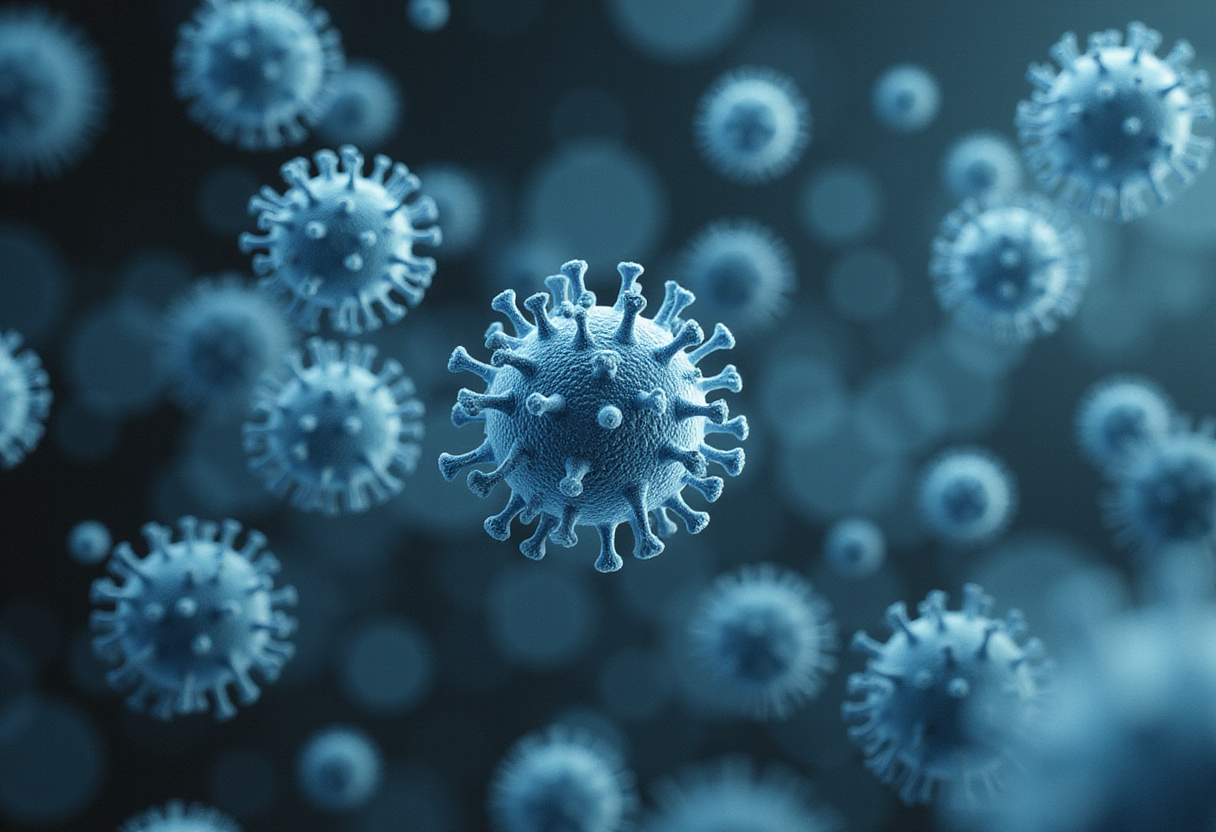
A study published Sept. 10 in Science has identified the exact protein that lets rotavirus punch through a cell membrane — and it's not the one researchers had bet on for years.
The team, led by Harvard Medical School research fellow Marilina de Sautu, showed that a protein called VP7, not VP5, forms the pore that lets the virus push its RNA into a cell. Stephen Harrison, senior author and the Giovanni Armenise-Harvard Professor of Basic Medical Sciences at HMS, had spent decades expecting the opposite answer.
"All along I had thought it would be VP5 that punched the hole, but it's VP7," Harrison said. "This turns out to be hole punching of a particularly satisfying type."
Why does one virus class matter so much?
Viruses come in two flavors, as Harrison likes to quip — not chocolate and vanilla, but enveloped and nonenveloped. The first group, which includes influenza, HIV, herpesvirus and SARS-CoV-2, wears a fatty membrane shell. Scientists understand those viruses' entry tricks fairly well.
The second group — poliovirus, HPV, rotavirus and adenovirus among them — carries its genome in a bare protein coat with no membrane disguise. Until now, researchers knew less about how these so-called naked viruses smuggle their genetic material across cellular barriers.
That gap matters beyond infection biology. "This work is relevant not just for understanding infection by nonenveloped viruses but for addressing the general question of what are the mechanisms for delivering large cargo into cells," said Harrison, who is also a Howard Hughes Medical Institute investigator. The findings could help biotech companies design better ways to ferry gene therapies and other bulky therapeutic payloads into cells.
How does the virus actually get in?
Previous work from the Harrison Lab and others had established the opening scenes of the story. A rotavirus attaches to the cell membrane, then the cell swallows it into a membrane-bound compartment — a kind of cellular waiting room.
The new study, which used a strain of rotavirus, filled in the rest of the script:
- Virus protein 5 (VP5) makes the compartment's membrane leaky, letting calcium ions escape
- The calcium loss causes virus protein 7 (VP7) to detach from the virus particle
- Freed VP7 then assembles into a pore in the membrane
- The virus pushes its RNA through that opening, hijacking the cell's machinery to replicate
Harrison credited earlier hints to Philip Dormitzer, a former HMS and Boston Children's colleague now a biopharmaceutical executive, and their student Shane Trask, whose work had pointed toward VP7.
What made the molecular movie possible?
The technique at the heart of the paper is cryo-electron tomography, or cryo-ET. Think of it as a CT scan for molecules: researchers flash-freeze an ultrathin sample, snap high-resolution images from many angles, and reconstruct them into a 3D model. The difference is that the microscope stays still while the sample tilts, rather than the device circling the patient.
The reconstructions captured every step of rotavirus infection — attachment, engulfment, membrane puncture and RNA ejection. Richard Walsh and Conny Leistner at the Harvard Cryo-EM Center for Structural Biology at HMS, which Harrison directs, supported the imaging work. Live-cell and single-molecule imaging from coauthor Tomas Kirchhausen's lab helped motivate and validate the findings.
"Cryo-ET allowed us to do something we wouldn't have been able to do even a few years ago," Harrison said. He noted that a paper by HMS professor Alan Brown on cilia, published online in Science the same day, shows the technology's growing reach.
What's still uncertain?
The results are preliminary on one front: an important set of experiments remains to confirm the fine details of how VP7 builds its pore, Harrison cautioned.
The team's next moves are already mapped. Kirchhausen has laid groundwork for imaging the entry steps of adeno-associated viruses, or AAVs — the workhorse delivery vehicles that pharma companies commonly use for gene therapies. De Sautu plans to keep probing viral entry as she finishes her postdoctoral work and starts her own lab.
"Curiosity about how evolution has solved the problem of delivering a large particle into a cell without harming it is my principal motivation," Harrison said, "but I'm thrilled that it has potential implications from which biotechnologists can take inspiration."
The study appears in Science (2026), DOI: 10.1126/science.aeg4851.
via Phys.org Biology (Source)
More from Nathan Brooks
Show full bio
Market editor covering companies, deals and strategy in general science news at SciBeat.
220 articles
Nearby plates
- COVID-19 immunity may shield against other bat coronaviruses
- Nasal Vaccine Additive Kept Mice Alive Against Flu 100 Days Later
- MIT team builds method that lets living cells mail out their own RNA
- MIT Automates Lipid Nanoparticle Production for RNA Therapies
- Oxford Team Watches Virus-Like Particles Build Themselves in Real Time