Plate Nº 24 · recorded October 10, 2026
Neuroscience & MindReported finding
Why SCA1 Destroys the Cerebellum but Spares Other Brain Regions
A new Baylor College of Medicine study in Genes & Development explains why mutant ATXN1 damages only the cerebellum: its preferred partner protein is most abundant there.
By Priya Raman4 min read817 words
In brief
- Mutant ATXN1 protein is expressed throughout the body, yet SCA1 primarily destroys the cerebellum and brainstem
- CIC-Long preferentially binds ATXN1; CIC-Short preferentially binds ATXN1L, and the two forms are not interchangeable
- Mice lacking CIC-S suffered lung defects, hydrocephalus and early death; mice lacking CIC-L survived with memory and movement problems
- The cerebellum carries the highest CIC levels in the central nervous system, explaining its vulnerability
- Findings appear in Genes & Development, from Baylor College of Medicine and the Duncan NRI at Texas Children's Hospital
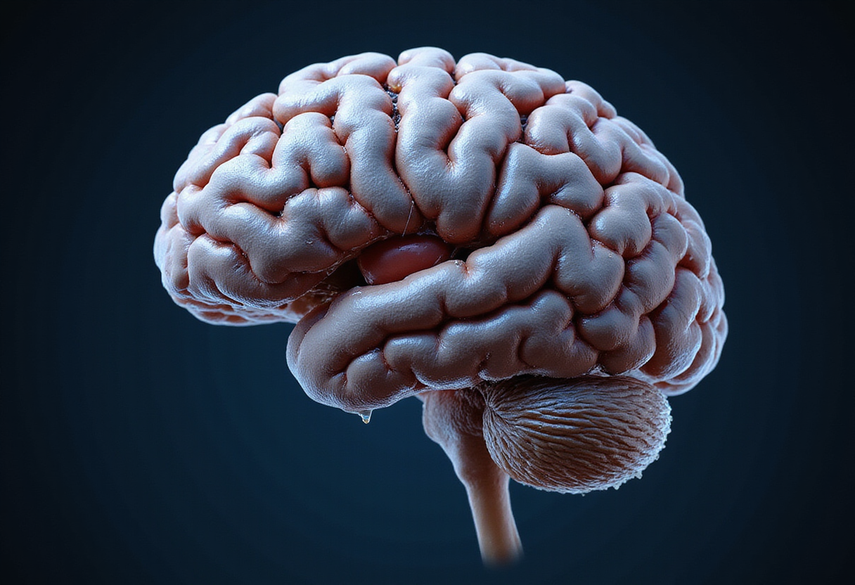
The mutant protein behind spinocerebellar ataxia type 1 exists throughout the human body — in the heart, the liver and most brain regions — yet it destroys mainly the cerebellum and brainstem. Researchers at Baylor College of Medicine and the Duncan Neurological Research Institute (Duncan NRI) at Texas Children's Hospital now explain why, in a study published in Genes & Development.
Their answer lies not in the mutant protein itself but in its partners. Different tissues carry different amounts of two forms of a partner protein called Capicua (CIC), and the mutant protein preferentially grabs one of them. Where its favorite partner is most abundant, damage concentrates.
What is SCA1?
SCA1 is a rare neurodegenerative disorder caused by a mutation in the ATXN1 gene. The mutation produces a faulty protein that is overly active and accumulates inside cells, eventually killing them.
"SCA1 is a rare neurodegenerative disorder characterized by progressive loss of coordination (ataxia), slurred speech and swallowing difficulties, which result from damage to the cerebellum, the brain region that controls coordination and balance," said corresponding author Dr. Huda Zoghbi, Distinguished Service Professor at Baylor, director of the Duncan NRI and a Howard Hughes Medical Institute investigator.
That selectivity has long puzzled researchers. If the defective protein is everywhere, why do only certain tissues degenerate?
How did the team tackle the puzzle?
The researchers, led by first author Hamin Lee, a graduate student in the Zoghbi lab, worked with genetically engineered mouse models and went back to protein basics.
Earlier work had shown that ATXN1 stabilizes a partner protein, CIC, which is also expressed throughout the body. CIC comes in two forms: CIC-Long (CIC-L) and CIC-Short (CIC-S). The two differ at one end of the protein, hinting at different jobs.
"We knew that CIC exists in two forms, CIC-Long (CIC-L) and CIC-Short (CIC-S)," Lee said. "Both ATXN1 and ATXN1L bind to the same section on both CICs. CIC-L and CIC-S differ at one end of the protein, suggesting that the two forms play distinct biological roles."
The body also produces a cousin of ATXN1 called ataxin-1-like (ATXN1L). Losing each protein in mice produces very different outcomes:
- Losing ATXN1 does not cause ataxia. Instead, it produces learning and memory deficits and increases amyloid beta production — Alzheimer's-like features affecting the cortex and hippocampus.
- Losing ATXN1L causes lung defects, death of newborns shortly after birth, and hydrocephalus, an abnormal buildup of cerebrospinal fluid in the brain's cavities.
What did the mice reveal?
When the team engineered mice to lack one CIC form at a time, the differences were striking:
- Mice without CIC-S often died early, with severe developmental problems especially in the lungs. Some also accumulated fluid in the brain, grew poorly and eventually died.
- Mice without CIC-L survived but showed behavioral abnormalities, learning and memory difficulties, movement deficits and hyperactivity.
"These findings showed that the two forms of CIC are not interchangeable, each has its own essential function," Lee said.
The symptom overlap was telling: mice lacking CIC-S resembled mice lacking ATXN1L, and mice lacking CIC-L resembled mice lacking ATXN1. Closer examination confirmed specific pairings — CIC-L preferentially binds ATXN1, while CIC-S preferentially binds ATXN1L.
Why does the cerebellum take the hit?
Protein levels vary by brain region and developmental stage. The cerebellum carries the highest baseline levels of CIC in the central nervous system. When overactive mutant ATXN1 arrives, it finds a dense pool of its preferred CIC-L partners there, hyper-stabilizing the complex and disrupting gene regulation specifically within cerebellar Purkinje networks. Other tissues, with lower CIC-L ratios, are spared.
"This means that certain areas are more vulnerable if a specific protein partnership is disrupted," Lee said.
During lung development, ATXN1L and CIC-S are both at high levels, which mirrors the lung failure seen when that pairing is broken.
"Our findings show that subtle differences in the relative abundance of CIC and ATXN1 forms at the protein level, together with the differential complexes they assemble, can result in highly specialized functions and dictate regional vulnerability," Zoghbi said.
What could this mean for treatment?
The findings come from mouse models, so human applications remain preliminary. Still, the study suggests a practical strategy: instead of shutting down a disease protein everywhere and risking side effects in healthy tissue, future therapies could target the specific protein partnership driving disease in a given region — such as the ATXN1/CIC-L complex in the cerebellum.
"Our study illuminates an improved understanding of neurological disease and offers new possibilities to comprehend and treat these conditions more effectively," Zoghbi said.
Esmeralda Villavicencio Gonzalez, Elias M. Rivera, Mark A. Durham, Ronald Richman, Elizabeth H.-Y. Chu, Kailey Xia, Hu Chen, Zhandong Liu, Surabi Veeraragavan and Binoy Shivanna, all at Baylor College of Medicine and/or Texas Children's Hospital, also contributed to the work.
via utoronto.ca (Original)
More from Priya Raman
Show full bio
Senior reporter covering industry trends and analytics at SciBeat.
207 articles
Nearby plates
- Doubling Lifespan in Parkinson's Mice: How One Protein Stopped Toxic Clumps
- Phase 3 Results for Oral Fabry Therapy Lucerastat Published in Nature
- MSK Reports Early Promise for KRAS-Targeted Pancreatic Cancer Therapy
- Roche Halts Two Huntington's Disease Drug Programmes
- Three Cell Clusters Drive Muscle Wasting in Pancreatic Cancer