Plate Nº 86 · recorded November 13, 2026
Neuroscience & MindReported finding
The Brain's Fear Center Doubles as a Save Button for Memory
Neurons in the amygdala, the brain's fear hub, predicted which images 90-plus patients later recalled, a Nature Neuroscience study reports. The region may help sort what we save.
By Nathan Brooks3 min read645 words
In brief
- A study published October 27 in Nature Neuroscience links individual amygdala neurons to memory formation.
- Researchers recorded single-neuron activity in more than 90 people with drug-resistant epilepsy.
- Patients viewed images in five categories, then tried to recall them 10-30 minutes later.
- Amygdala neuron responses on first viewing predicted later recall; the hippocampus signal was weaker and delayed.
- No broadly effective treatments for memory disorders currently exist, Xiao notes.
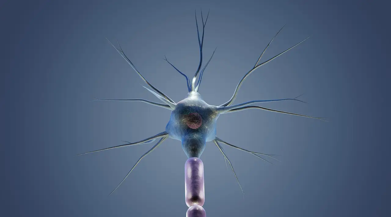
Neurons deep in the amygdala — the brain's walnut-sized fear hub — predict which images a person will remember, according to a study published October 27 in Nature Neuroscience. Activity in certain amygdala neurons, recorded at the single-cell level in more than 90 people, tracked whether an image was later recalled. That's a first for a region usually cast as the brain's emotion regulator, not its filing clerk.
The finding chips away at a basic puzzle of the mind. "We forget much of what we experience … only a fraction of the information we encounter becomes a lasting memory," says Jiayang Xiao, a neuroscientist at Cedars-Sinai Health Sciences University in Los Angeles and one of the study's authors.
How did researchers watch single neurons?
Earlier work had hinted the amygdala plays some role in memory, but the tools were coarse. Electrodes on the scalp, or arrays reading thousands of neurons at once, produced what Cory Inman calls the stadium effect. Inman, a cognitive neuroscientist at the University of Utah in Salt Lake City who was not involved in the study, describes it this way: recording from outside a stadium, "you might be able to tell that there's a game happening" — a touchdown, maybe, or which side of the crowd is cheering. That's about it.
The new study is more like a lapel mic pinned on a single fan. Xiao and her colleagues used electrodes already implanted in the brains of more than 90 people with drug-resistant epilepsy, placed there to locate where seizures start. Borrowing that clinical hardware, the team eavesdropped on individual amygdala neurons — a region that's hard to reach because it sits in two clusters deep inside the brain.
What did the patients actually do?
The task was simple:
- Patients viewed images of everyday objects from five categories, such as food and animals.
- Ten to 30 minutes later, they tried to recall which images they had seen.
- Researchers compared each neuron's activity at first viewing with later recall.
The result: activity in certain amygdala neurons corresponded with an image being remembered. In effect, the image had been earmarked for longer-term storage, Xiao says.
What's the proposed mechanism?
The study doesn't nail down exactly how a memory gets saved, and the researchers are careful about that. But their data suggest a two-step process. First, the amygdala categorizes the image — individual neurons showed a preference for one of the five image categories. Second, the strength of that category neuron's response on the very first viewing dictates whether the image is later recalled.
There are real limits to keep in mind. Participants only had to remember things from half an hour earlier at most, so the findings may not extend to memories that last days or years. And this was a group of epilepsy patients with implanted electrodes, not a random sample of brains.
The surprise, Xiao says, is where the signal showed up. The hippocampus is the region most directly linked to memory, so "it surprised me that we observe such a clear relationship between memory formation and activity in the amygdala." By contrast, the link between hippocampus neuron activity and image recall was weaker, and it arrived slightly later.
Why does this matter?
A clearer picture of this mechanism could eventually guide better treatments for memory disorders — a medical problem with no broadly effective options today. That prospect is distant, and Xiao frames it cautiously. The next step, she says, is work like stimulating amygdala neurons and observing the impact on memory, which would tell researchers whether this region can actually be targeted to improve recall.
For now, the machinery behind what we remember remains partly in the shadows. But the amygdala just earned a promotion: from the brain's fear center to a key clerk in its filing system.
via sciencenews.org (Source)
More from Nathan Brooks
Show full bio
Market editor covering companies, deals and strategy in general science news at SciBeat.
338 articles
Nearby plates
- Scientists reconstruct 10-second videos from mouse brain signals
- The Unconscious Brain Predicts Words, Baylor Study Finds
- Mouse Study Maps a Brain Circuit That Tunes Stress Responses
- Fruit Fly Brains Reveal How a Neural Gate Switches Memory On
- Rutgers study: fear boundaries form in early brain sensory circuits