Plate Nº 61 · recorded October 10, 2026
Neuroscience & MindReported finding
Human Brain Grows From Two Separate Cell Lineages, Study Finds
Stanford scientists discovered the brain forms from two independent cell lineages, a 550-million-year-old design that enabled the first lab-grown human hindbrain neurons for ALS and SMA research.
By Marcus Bennett4 min read767 words
In brief
- The human brain develops from two separate, non-overlapping progenitor lineages — Otx2+ for forebrain/midbrain and Gbx2+ for hindbrain — not one common progenitor.
- The same dual-origin mechanism appears in species that diverged from our lineage over 550 million years ago.
- Researchers grew functional, electrically active human hindbrain motor neurons in the lab for the first time.
- The study was published in Nature Neuroscience on September 18, 2026, by a team at Stanford Medicine.
- Distinct chromatin packaging locks each lineage onto its developmental track, explaining decades of failed attempts to convert forebrain precursors into hindbrain tissue.
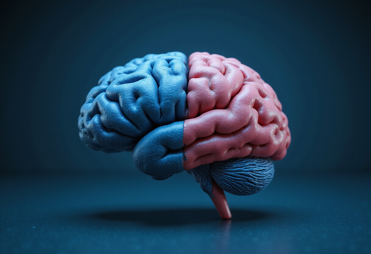
For the first time, Stanford Medicine researchers have grown authentic human hindbrain neurons in the laboratory — a feat made possible by their discovery that the vertebrate brain does not descend from a single progenitor cell, but from two distinct, mutually exclusive lineages that evolution joined side by side more than 550 million years ago.
The study, published in Nature Neuroscience, refutes a foundational assumption that has stood in anatomy textbooks for centuries: that one common pool of embryonic progenitor cells diversifies into all regions of the nervous system.
"We've shown for the first time that the front of the brain arises from a totally different progenitor cell than the back of the brain," said Kyle Loh, Ph.D., associate professor of developmental biology at Stanford Medicine and senior author of the study. "Our discovery means that we can now grow neurons from the back of the brain, the hindbrain, in a petri dish and study their functions."
What did the researchers find?
Co-first authors Rayyan Jokhai and Carolyn Dundes traced cell fates in mouse embryos during gastrulation — the earliest phase of embryonic development, when the body's basic layout takes shape. They identified two completely non-overlapping progenitor populations:
- Anterior lineage (Otx2+): destined exclusively to build the forebrain and midbrain, the seat of executive cognition, language, and abstract thought.
- Posterior lineage (Gbx2+): committed from the outset to constructing the hindbrain, or brainstem, which controls breathing, heart rate regulation, sleep-wake cycles, and motor control over facial expression, speech, and swallowing.
The two populations never cross lineages. Epigenomic analysis — a readout of how DNA is packaged inside cells — showed that the anterior and posterior neural ectoderm carry fundamentally distinct chromatin configurations, the physical packaging of genetic material that locks each cell type onto its developmental track from the very start.
This locked-in packaging explains a long-standing laboratory failure. "Previous attempts to make hindbrain neurons likely tried to coax forebrain and midbrain progenitors into hindbrain cells, which our study shows is not possible," Jokhai explained. "In stem cell biology, people are always fixated with creating the end cell type, like the neuron. But it's important to begin at the earliest stages of embryonic development. Our careful attention to that early time point allowed us to find this fundamental split."
How old is this dual-brain design?
To trace the divide's evolutionary history, the team examined animals across distant branches of the tree of life. They found the same dual-origin mechanism in chickens, zebrafish, and marine acorn worms, which split from our shared lineage over 550 million years ago. Even more primitive organisms, such as jellyfish that diverged 600 to 700 million years ago, carry two separate nervous systems at opposite ends of their bodies.
"Our research suggests that evolution took two existing neural systems and pushed them together spatially," Loh said. "Having the brain as one organ would probably be more efficient, but we rely on this primordial way to make the brain as two separate pieces."
Why does this matter for disease research?
By following the natural developmental split rather than fighting it, the Stanford team directed human pluripotent stem cells — master cells capable of becoming any tissue — into fully functional hindbrain motor neurons. The lab-grown cells fired normal electrical signals and expressed markers of hindbrain segments that control facial muscles and swallowing.
The breakthrough opens a long-missing experimental platform. In disorders such as amyotrophic lateral sclerosis (ALS) and spinal muscular atrophy (SMA), degenerating hindbrain motor neurons rob patients of the ability to swallow and breathe, often leading to fatal aspiration pneumonia or respiratory arrest. Researchers cannot take living brainstem tissue from patients, so the field has lacked accessible human cellular models — until now.
The system may also inform metabolic medicine. The hindbrain houses the neural circuits that regulate hunger and satiety — the very pathways targeted by GLP-1 receptor agonists such as semaglutide. Lab-grown hindbrain tissue will let pharmacologists directly test how appetite-suppressing drugs interact with human brainstem circuits.
"Now we have a model to better understand these devastating diseases, and work toward regenerative therapies for them," Jokhai said. "This is a very exciting new frontier in brain research."
What are the caveats?
The core lineage-tracing results come from mouse embryos, and the evolutionary argument rests on comparisons across chickens, zebrafish, and acorn worms. The paper proposes — but has not proven — that the dual-progenitor design is conserved across all vertebrates. The stem-cell-derived hindbrain neurons are a research model, not a therapy, and any clinical application for ALS or SMA remains years away.
via stanford.edu (Original)
More from Marcus Bennett
Nearby plates
- Brain Arises From Two Separate Cell Sources, Not One, Study Finds
- Metabolism and Touch: Two Hidden Forces That Shape the Human Brain
- Brain Wiring Starts Precise, Not Random, Mouse Study Finds
- Chameleon Embryos Share a Developmental Trick Once Thought Unique to Mammals
- No Lab-Grown Embryo Model Fully Matches the Real Thing, Study Finds