Plate Nº 29 · recorded October 10, 2026
Biology & EvolutionReported finding
No Lab-Grown Embryo Model Fully Matches the Real Thing, Study Finds
A Sydney team benchmarked four stem-cell-derived blastoid models against a map of over 14,000 single embryonic cells. Some scored well; none matched the real thing.
By James Calloway3 min read672 words
In brief
- University of Sydney researchers evaluated four leading blastoid-generation protocols and found none perfectly replicated a natural human blastocyst.
- The study, published in Cell Systems (2026), is led by Associate Professor Pengyi Yang.
- The team built a reference map from more than 14,000 single-cell transcriptomes from human embryos.
- The reference datasets and benchmarking tools are publicly available to researchers worldwide.
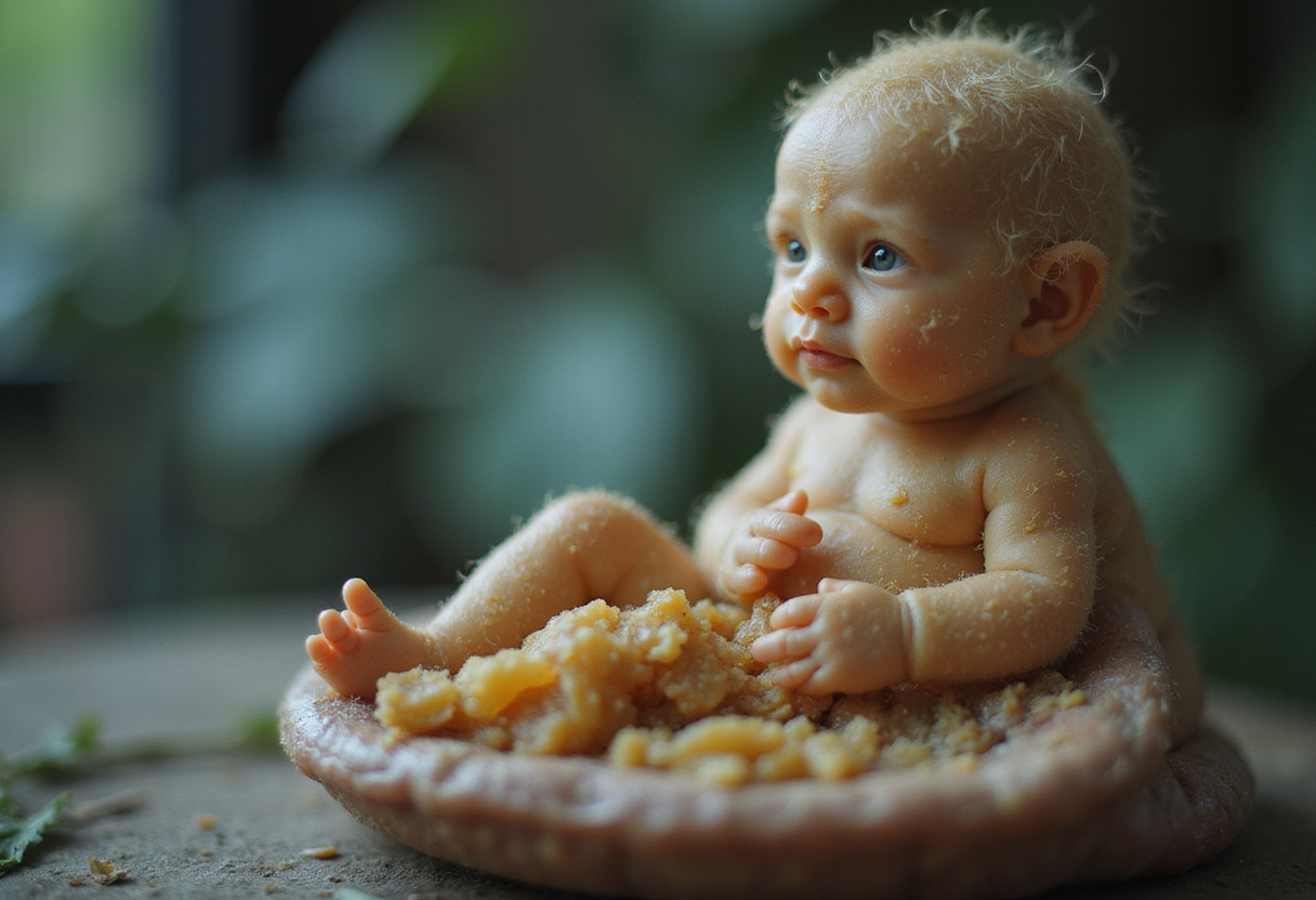
None of the four leading lab-grown models of the early human embryo accurately reproduces every cell type found in a natural one, according to a University of Sydney study published in the journal Cell Systems. The work offers the field what its authors call an objective "reality check": some models perform well, but none yet captures the full complexity of early human development.
The models in question are called blastoids — structures grown from stem cells that mimic the blastocyst, the tiny ball of cells an embryo forms in the days before and after it implants in the uterus. Researchers use blastoids to study fertility, pregnancy success and the earliest stages of human development without relying on donated embryos, which are scarce and raise ethical concerns.
Importantly, blastoids are research tools, not embryos. Current models cannot develop into a human being.
What did the researchers actually build?
The team, led by Associate Professor Pengyi Yang of the School of Mathematics and Statistics and the Children's Medical Research Institute, developed a computational framework that tests how closely a blastoid behaves like a real embryo at the molecular level — not just how much it looks like one under a microscope.
To do this, they combined and harmonized more than 14,000 single-cell transcriptomes — snapshots of the RNA molecules inside individual cells — taken from human embryos at key developmental stages. The result is one of the most comprehensive reference maps yet of how cells normally specialize and organize themselves in the days surrounding implantation.
The researchers then benchmarked four widely used blastoid-generation protocols, developed by international groups, against that map. They compared molecular identities, developmental timing and lineage structure rather than appearance alone.
How did the models score?
The evaluation revealed substantial differences between methods:
- Some blastoids reproduced all three major cell lineages of a natural human blastocyst relatively well.
- Others misrepresented certain cell types entirely.
- Some models contained large numbers of cells that could not be confidently matched to any known embryonic state.
- No single model perfectly replicated a natural human blastocyst.
"The encouraging finding is that some models capture important aspects of early embryonic development relatively well, although each model has limitations," Yang said.
Why does this matter?
Before this study, the field lacked a consistent yardstick. Looking embryo-like under a microscope says little about whether a model's inner biology matches the real thing.
"Human embryo models have enormous potential for studying the earliest days of an embryo's development, but there has been no consistent way to assess how accurately these reflect real human development," said Yang, who also leads the Trans-Regulatory Biology group at the Charles Perkins Center.
"Our framework allows researchers to compare these models against a detailed biological reference and determine which cell types and developmental processes are faithfully reproduced, and which are not."
The findings come with a clear caution. "Our study also shows that current models are not biologically equivalent to real human embryos, and researchers need to be careful about the conclusions they draw from them," Yang said.
What happens next?
The researchers have made their reference datasets and benchmarking tools publicly available, so scientists anywhere can test new embryo models against the same standards. They hope a shared benchmark will speed up improvements and make evaluations more rigorous across the field.
"If we're going to use these systems to answer important biological questions, we first need to know what they can reliably tell us," Yang said. "Our work provides a roadmap for improving embryo models and ensuring scientific claims remain grounded in what the models can actually support."
The study, by Long, S. et al., appears in Cell Systems (DOI: 10.1016/j.cels.2026.101738).
As with any benchmark, this one reflects the embryo data currently available; the map covers a limited window of early development. But as a first standardized test of its kind, it gives researchers a concrete way to separate what blastoids can faithfully tell us about human beginnings from what they cannot — yet.
via Phys.org Biology (Source)
More from James Calloway
Show full bio
Staff writer covering marketplaces and e-commerce at SciBeat.
205 articles
Nearby plates
- Chameleon Embryos Share a Developmental Trick Once Thought Unique to Mammals
- Scientists Build the First Million-Cell Family Tree of a Developing Mouse
- Human Brain Grows From Two Separate Cell Lineages, Study Finds
- Brain Arises From Two Separate Cell Sources, Not One, Study Finds
- MIT team builds method that lets living cells mail out their own RNA