Plate Nº 49 · recorded October 7, 2026
Biology & EvolutionReported finding
Two Protein Plugs Help Superbug P. aeruginosa Block Antibiotics
Two plug proteins, SlkA and SlkB, seal the outer membrane channels of P. aeruginosa against antibiotics, a 2026 Nature Communications study reveals — offering a new drug target.
By Nathan Brooks3 min read692 words
In brief
- A 2026 Nature Communications study identified two proteins, SlkA and SlkB, that plug antibiotic-entry channels in P. aeruginosa.
- Researchers from Sungkyunkwan University and The Catholic University of Korea, led by Professors Hongbaek Cho and Jeong Min Chung, made the discovery.
- Cryo-electron microscopy revealed high-resolution 3D structures of the plug proteins bound inside the channel.
- The plugs seal the type IV pilus assembly channel before the pili are fully formed, keeping the outer membrane barrier intact.
- The mechanism may serve as a target for next-generation antimicrobials and adjuvant therapies against gram-negative bacteria.
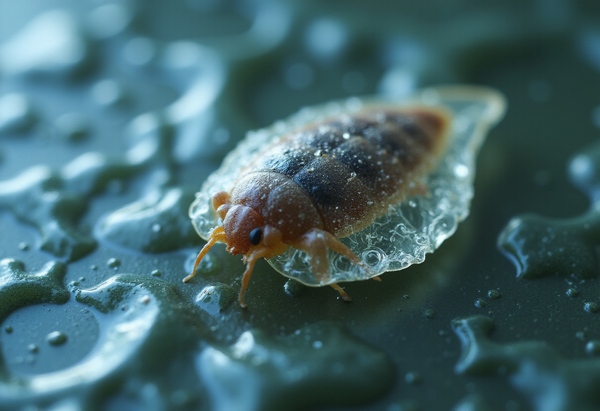
Two proteins act as physical plugs that seal the channels of the superbug Pseudomonas aeruginosa against antibiotics, according to a study published in Nature Communications in 2026. The finding reveals a previously unknown resistance mechanism and points to a possible new target for drugs against multidrug-resistant bacteria.
A joint research team led by Professor Hongbaek Cho of the Department of Biological Sciences at Sungkyunkwan University (SKKU) and Professor Jeong Min Chung of the Department of Biotechnology at The Catholic University of Korea made the discovery. Using cryo-electron microscopy, a technique that freezes molecules in place to capture sharp three-dimensional images, they visualized the plug proteins bound inside the bacterial channel at high resolution.
Why is P. aeruginosa so hard to kill?
Antibiotic resistance in pathogenic bacteria has become a major global public health threat. Multidrug-resistant bacteria, often called "superbugs," can survive treatment with multiple antibiotics, which makes infections extremely difficult to treat.
P. aeruginosa is one of the most notorious members of this group. It causes healthcare-associated infections, pneumonia, sepsis and other serious conditions, particularly in hospital settings.
Part of its resilience comes from its outer membrane — a tough outer wall that blocks antibiotics from penetrating the cell. But the bacterium faces a design problem of its own. To infect a host, it must build threadlike surface structures called type IV pili (T4P), which help it attach to host cells. Assembling those pili requires channels that pass through the protective outer membrane.
Until now, researchers did not understand how P. aeruginosa keeps those infection-critical channels open for business while still shutting antibiotics out.
What did the researchers find?
The answer, the team discovered, is a pair of dedicated gatekeepers. Two proteins, named SlkA and SlkB, bind inside the channel of the T4P assembly machinery and act as physical plugs.
The timing matters. The plugs do their job during a vulnerable stage before T4P is fully assembled, when the channel would otherwise leave a gap in the outer membrane. By sealing that gap, the proteins maintain the integrity of the membrane barrier and prevent antibiotic molecules from flowing into the cell.
Using cryo-electron microscopy, the researchers captured high-resolution three-dimensional structures of both proteins bound within the channel. These images show exactly how the plugs fit inside the pore.
The discovery overturns an earlier assumption. Scientists previously believed the channel itself was shaped in a way that blocked antibiotic entry. The new study shows a different mechanism: dedicated proteins actively plug the hole and reinforce the outer membrane's protective function.
The research team explained that the finding resolves a long-standing puzzle — how the bacterium can maintain structures essential for infection while preserving its defense against antibiotics.
Could this lead to new treatments?
The researchers see therapeutic potential in the mechanism itself. If a drug could disrupt the plug proteins, or prevent them from binding, antibiotics might slip through the outer membrane during the vulnerable assembly window.
The team said the discovery could provide a foundation for developing next-generation antimicrobial agents and adjuvant therapies — drugs given alongside antibiotics to boost their effect — that target the outer membrane barrier of gram-negative bacteria, the broad class of microbes with this type of double-membrane armor.
By disrupting the mechanism that keeps antibiotics out, the findings may open new strategies for overcoming multidrug resistance in P. aeruginosa specifically.
As with any single study, translation from mechanism to medicine will take time. The results are early-stage, and the researchers have so far characterized the plugs' structures and function rather than testing drugs against them. Whether compounds can be designed to pull the plugs in a living infection remains an open question for future work.
Still, the study offers something valuable for a field in urgent need of new targets: a precise, visualized picture of how one superbug defends itself — and, by extension, where its defenses might be broken.
Publication details
The study, "Secretin-interacting plug proteins prevent antibiotic influx during type IV pilus assembly in Pseudomonas aeruginosa," by Oh Hyun Kwon et al., appears in Nature Communications (2026), DOI: 10.1038/s41467-026-73864-w.
via Phys.org Biology (Source)
More from Nathan Brooks
Nearby plates
- Scientists Map Three Weak Points in Antibiotic-Resistant Bacteria
- Resistance Gene Helps C. difficile Survive Hospital Disinfectants
- Scientists Find a Way to Kill a Drug-Resistant Deadly Fungus
- Why a Food Poisoning Bacterium Can Also Cause Severe Wound Infections
- Common Mouth Bacterium Trains Lungs to Fight Pneumonia, Study Finds