Plate Nº 10 · recorded November 17, 2026
Biology & EvolutionReported finding
UCLA Adds a Lid to Nanovials, Unlocking Longer Cell Experiments
UCLA researchers capped nanovials with hydrogel spheres, trapping bacteria, yeast and mammalian cells for up to three days and enabling parallel colony growth and cell-to-cell tests.
By Marcus Bennett4 min read829 words
In brief
- Up to 9 out of 10 nanovials self-assemble caps after process optimization, per the Science Advances study (2026).
- Capped nanovials confined bacteria for 6 hours, yeast for 12 hours and engineered mouse cells for 3 days.
- The cap uses vitamin B7 on the bowl rim and a bacterial protein on the cap, bonding like molecular Velcro.
- The platform runs on standard lab sorting equipment, not specialized instruments.
- First author Michael Mellody; senior author Dino Di Carlo, UCLA bioengineering chair and Partillion Bioscience co-founder.
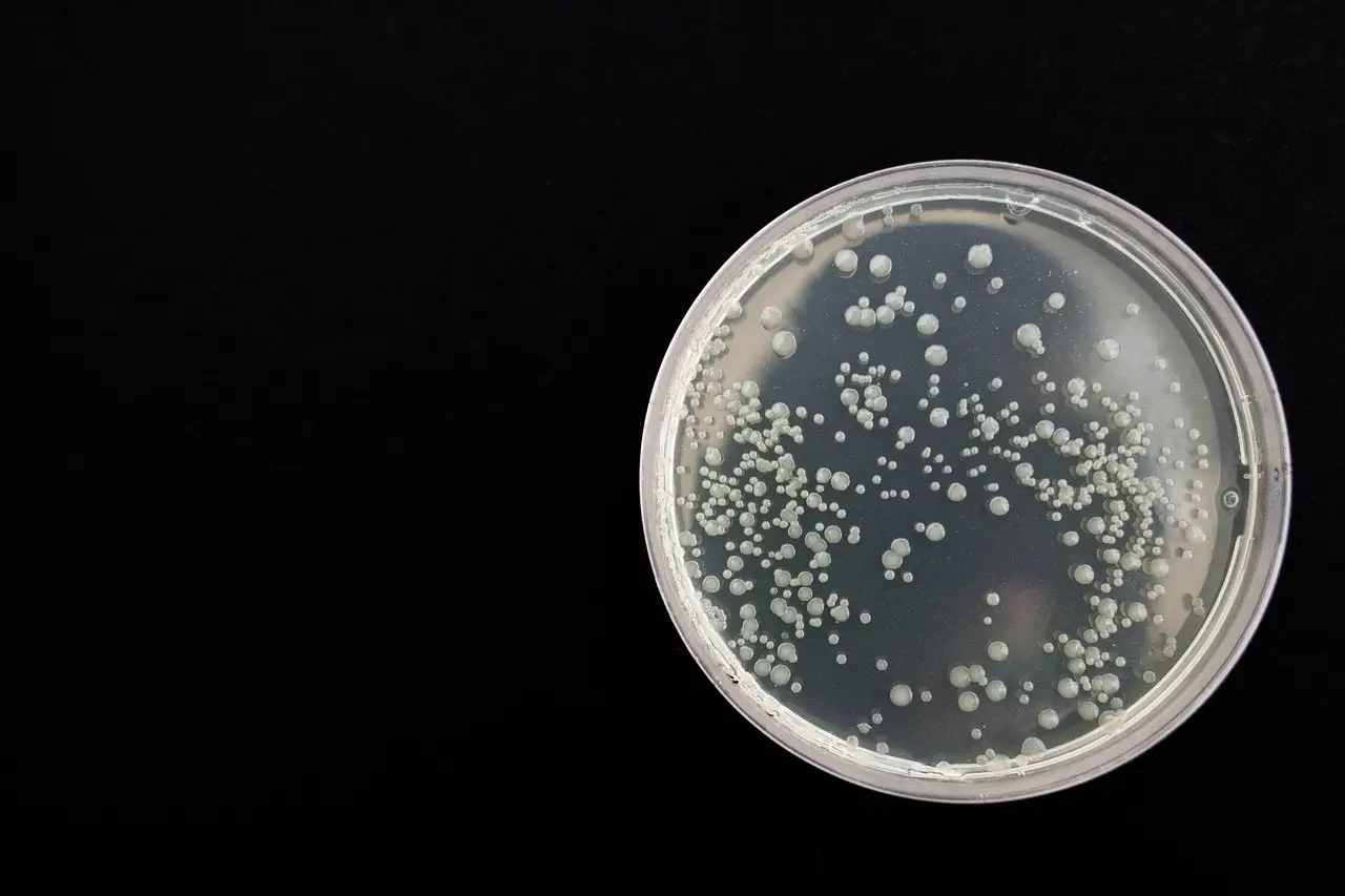
Up to nine out of 10 tiny hydrogel "test tubes" now seal shut at UCLA, and that lid turns a short-lived cell-trapping tool into a platform that can grow colonies for days and watch one cell change another's behavior. A study published in Science Advances on the technology, developed by Dino Di Carlo's lab at the UCLA Samueli School of Engineering, shows what becomes possible when cells stay put.
Nanovials are bowl-shaped hydrogel particles tens of micrometers across—imagine a test tube shrunk until it holds one or two cells, then manufactured by the hundreds of thousands so experiments run in parallel. The bowls capture cells and measure what those cells secrete. The catch: the trap was temporary. "On their own, nanovials compartmentalize cells and their products, but it's kind of short-term," said Di Carlo, the Armond and Elena Hairapetian Professor of Engineering and Medicine and chair of UCLA's bioengineering department. "The capping system opens up other types of longer-term experiments that involve growing colonies or selecting bacteria or yeast based on certain characteristics."
How do you cap half a million microscopic bowls?
The idea came from a glitch. In the previous generation of the technology, one nanovial would occasionally nest inside another's cavity and block it. "At first we said, 'Oh, that's a problem,' but then we asked, 'What if we turn it into a solution?'" said Di Carlo, who is also a member of the California NanoSystems Institute at UCLA.
The caps are small hydrogel spheres. Researchers decorated the bowl's rim with vitamin B7 and the cap with a bacterial protein—two molecules that bond like molecular Velcro. Then they spun bowls and caps together in a centrifuge and let them settle. The bowls landed first, cavities facing up; the caps fell into place. After optimization, self-assembly capped as many as nine in 10 nanovials.
Because both bowl and cap are porous, nutrients flow in and waste flows out while cells stay confined. In short: the lid keeps cells in, not chemistry out.
What does the cap change for practitioners?
Plenty, according to the study, led by first author Michael Mellody. Three gains stand out:
- New cell types. Uncapped nanovials catch mammalian cells through protein-binding coatings on their interior. Bacteria and yeast lack those surface proteins and drift out. Inside capped nanovials, they stayed confined without any attachment—no species-specific engineering required. "We don't need to find a particular way to capture each species, and that lowers the barrier for someone to apply this approach," said Di Carlo, a co-founder of Partillion Bioscience, which commercializes nanovials. "The caps move nanovials toward being a generalized platform that doesn't require tuning, the same way you can use a petri dish to look at any type of cell."
- Colony growth. In uncapped bowls, dividing cells spill over the rim. Capped nanovials held growing bacterial colonies for six hours, yeast for 12 hours and engineered mouse cells for three days before the colonies popped the lid. Researchers could then sort those minicolonies by growth rate.
- Cleaner secretion data. For cells that pump out large amounts of protein, the extra containment gave a clearer signal than open bowls.
The system also runs on standard lab equipment. Unlike many micro-scale cell-testing setups that demand specialized instruments, capped nanovials can be analyzed and sorted with machines most biology labs already own.
What did the cell-to-cell experiments show?
The team placed antibody-producing cells together with engineered T cells that glow green when the antibody activates them. Because the cap kept cells and their signals in one compartment, the researchers could identify exactly which antibody producers switched on the T cells—an approach that could speed the discovery of more effective therapeutic antibodies.
"We've shown proof of principle for functional drug discovery tests," Di Carlo said. "You could also imagine basic studies putting any two cells together and looking at how they change each other over time through contact and secretion."
The results remain early-stage: the study demonstrates feasibility, not clinical findings, and colony lifetimes cap out at hours to days depending on the species.
What comes next?
The researchers are now screening bacteria for antibiotic susceptibility inside the compartments and testing whether capped nanovials can handle experiments that change conditions on captured cells over time.
They also see a fit with artificial intelligence. Predicting which cells grow fastest, produce useful medicines or respond to treatments requires massive experimental datasets—and each nanovial is one experiment. "These are very data-hungry systems, and it takes huge, high-quality data sets to create generalizable models," Di Carlo said. "We can't do five experiments in a day anymore. We have to do 100,000 experiments in a day."
Publication: Michael Mellody et al., "Massively parallel self-assembly of suspendable microcompartments for functional cell screening," Science Advances (2026). DOI: 10.1126/sciadv.aei7819.
via phys.org/rss-feed/ (Source)
More from Marcus Bennett
Show full bio
News editor covering regulation, policy and their impact on general science news at SciBeat.
450 articles
Nearby plates
- Superconducting Vortices Prove Clogging Is a True Phase Transition
- Kimchi Bacterium Trapped 87% of Nanoplastics — In a Lab Beaker
- Lab Chip With 444 Tiny Chambers Reproduces Herd Immunity
- Flexible molecular links create new nanoparticle crystals
- Oxford Team Watches Virus-Like Particles Build Themselves in Real Time