Plate Nº 86 · recorded November 11, 2026
PhysicsReported finding
Math Method Makes X-ray Images Three Times Sharper at Nanoscale
A new optimization method tripled 3D X-ray image resolution, resolving 20-nanometer structures like synapses without cutting or damaging samples, researchers report in Optica.
By Elena Vasquez3 min read610 words
In brief
- The new method resolves structures down to 20 nanometers; a human hair is 80,000-100,000 nanometers thick.
- Image resolution improved roughly threefold in the study, published in Optica in 2026.
- The technique enables 3D imaging of larger samples without slicing them, unlike electron microscopy.
- Marcus Carlsson of Lund University developed the optimization algorithm and is the paper's last author.
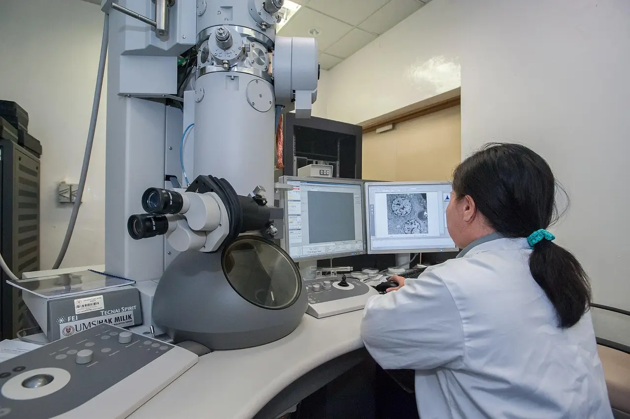
A new mathematical method has roughly tripled the resolution of 3D X-ray images, letting researchers pick out structures as small as 20 nanometers. For scale, a human hair measures 80,000 to 100,000 nanometers across. An international collaboration published the work in the journal Optica in 2026.
The technique targets X-ray nanotomography, a way of building three-dimensional images from X-ray scans. Until now, scientists largely blamed blurry results on the X-ray optics themselves. The new study says the math deserves part of the blame too.
"Our work shows that the limitation has not only been in the X-ray optics, but also in the mathematics used to reconstruct the image," said Marcus Carlsson, a mathematics researcher at Lund University and last author of the paper.
What got sharper?
The numbers tell the story. Image resolution improved about threefold with the new approach, according to the study. That jump brought very fine biological structures into view in three dimensions — synapses, mitochondria and other subcellular details inside the body's cells.
At that scale, resolution matters. Structures 20 nanometers wide sit deep inside the territory that biologists want to map, cell by cell.
Carlsson led the mathematical and algorithmic side of the research. He developed the optimization approach — in plain terms, a computational recipe that repeatedly refines an image until it best fits the measurement data — that underpins the refined imaging method.
Why not just use electron microscopes?
Researchers imaging the nanoscale usually reach for electron microscopy instead of X-rays. That tool delivers extremely high detail. But it comes with two costs.
First, samples must be cut into very thin slices for 3D imaging. Second, the process permanently damages the material being studied.
X-rays avoid both problems. They let scientists examine much larger samples at the nanoscale and image them in three dimensions without cutting anything apart. The trade-off has been resolution — until better math entered the picture.
Where does the math come in?
X-ray nanotomography generates enormous amounts of raw data. The hard part, Carlsson points out, is extracting all the information hiding inside those measurements. That's a computational problem, and it's where mathematics can pull weight that hardware alone cannot.
"With better mathematical models and optimization algorithms, we can extract significantly more information from the same type of measurement data," Carlsson said. "The new method opens up possibilities both in biological imaging and in other fields where researchers want to study materials three-dimensionally and nondestructively."
In other words, labs may not need new, more expensive X-ray equipment to see finer detail. Sharper software can squeeze more out of the scans they already collect.
What are the caveats?
The results come from a single study, and the threefold improvement reflects what the researchers measured in this work, not a guaranteed standard across all instruments and samples. Whether the method performs as well in everyday lab settings, across varied materials and sample types, will take further testing.
Still, the finding shifts some of the burden in imaging science. Better algorithms, not just better lenses, now look like a serious path to sharper pictures.
The paper's applications reach beyond biology. Any field that wants nondestructive 3D views of materials — think industrial components or layered electronics — could benefit from the same reconstruction approach.
The takeaway
Smarter math, applied to data scientists already collect, made X-ray images three times sharper and revealed cell structures just 20 nanometers across — no slicing, no destruction.
The published paper is Viktor Nikitin et al., "Scalable joint X-ray nano-holotomography reconstruction with the bilinear Hessian method," Optica (2026), DOI: 10.1364/optica.603920.
via phys.org/rss-feed/ (Source)
More from Elena Vasquez
Show full bio
Correspondent covering money, funding and market shifts around general science news at SciBeat.
384 articles
Nearby plates
- Microscope Upgrade Resolves 75 Nanometers Deep Inside Tissue
- Laser 'Nuisance' Speckle Powers Record 6-Femtometer Spectrometer
- A quantum computer is being wired into an electron microscope
- First near-atomic map of babesiosis parasite's ribosome unveiled
- AI and the Largest 2D Map of the Universe Uncover 70 New Gravitational Lenses