Plate Nº 22 · recorded November 11, 2026
Biology & EvolutionReported finding
Microscope Upgrade Resolves 75 Nanometers Deep Inside Tissue
Texas A&M researchers achieved 75-nanometer resolution deep in intact tissue by combining image scanning microscopy with optical fluctuation imaging on standard two-photon microscopes.
By Marcus Bennett3 min read613 words
In brief
- The 2PE-ISM-SOFI technique achieves lateral resolution of about 75 nanometers, roughly five times sharper than conventional two-photon microscopy.
- Findings were published in the Journal of Biomedical Optics in 2026, DOI: 10.1117/1.jbo.31.8.086501.
- Aart Verhoef's team at Texas A&M layered two established techniques onto a standard two-photon microscope.
- The study builds on groundwork published in PRX Life in 2024.
- Collaborators imaged midbrain dopamine neurons with tubulin labeled by quantum dots.
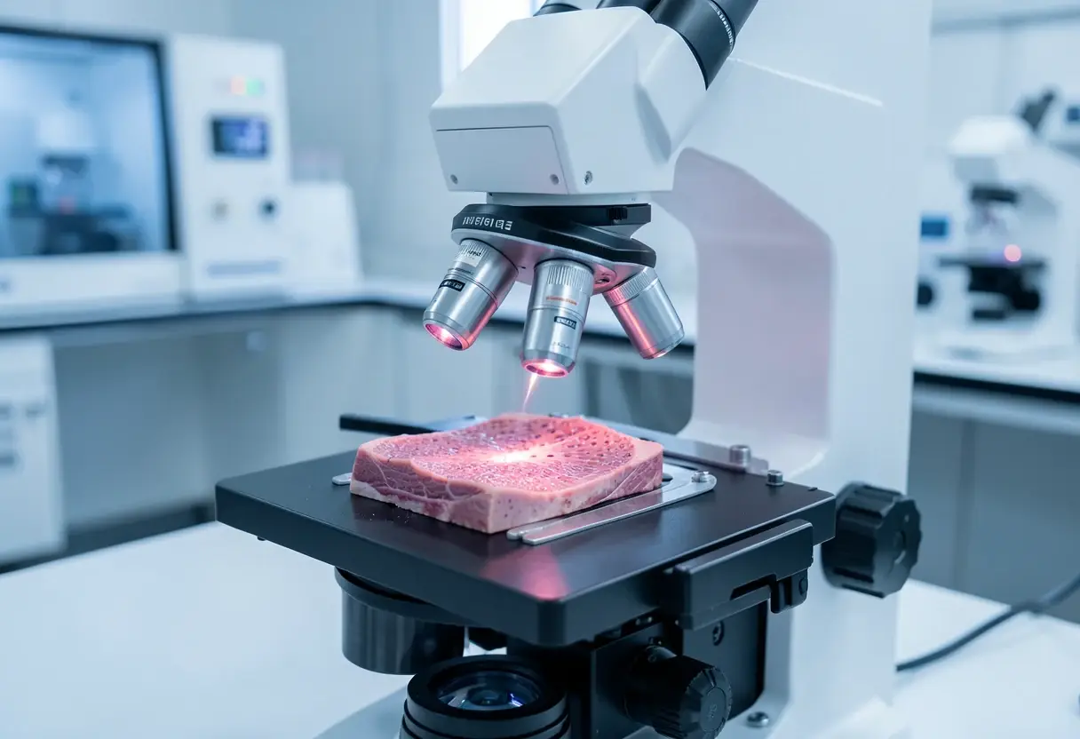
A new microscopy method resolves structures as small as 75 nanometers — roughly five times sharper than conventional two-photon microscopy — while keeping the deep-tissue imaging that biologists depend on. Researchers at Texas A&M University developed the technique and published their findings in the Journal of Biomedical Optics in 2026.
The method, called 2PE-ISM-SOFI, breaks a barrier that has constrained biological imaging for years: laboratories could either see fine cellular detail near a sample's surface or image deep into intact tissue, but rarely both at once. The new approach resolves structures smaller than 100 nanometers, about a thousand times thinner than a human hair, without sacrificing depth.
Aart Verhoef, Ph.D., assistant professor of biophotonics in the Department of Soil and Crop Sciences at the Texas A&M College of Agriculture and Life Sciences and joint professor in the Department of Physics and Astronomy, led the team.
"This technique builds on two microscopy techniques that were limited by depth and detail, but this technique allows us to look deep into tissue and improves the resolution," Verhoef said.
How does the upgrade work?
The researchers layered two established techniques onto a standard two-photon microscope rather than building new hardware from scratch. 2PE-ISM-SOFI stands for two-photon excited image scanning microscopy combined with super-resolution optical fluctuation imaging.
Two-photon microscopy earned its place in labs because it peers deep into biological samples with minimal damage — a requirement for studying living tissue in neuroscience, developmental biology, and soil-, animal- and plant-related sciences. The trade-off has always been resolution: fine detail typically meant staying near the surface.
Super-resolution techniques such as stimulated emission depletion (STED) and photoactivated localization microscopy (PALM) — work that earned their developers the 2014 Nobel Prize in Chemistry — can resolve features far below 100 nanometers, but usually only close to the surface. The new method holds on to two-photon imaging's depth advantage while closing in on that sharper resolution.
The takeaway: labs can now push toward sub-100-nanometer resolution on a standard two-photon platform without abandoning deep samples.
What did the team actually image?
Collaborators in the Texas A&M College of Medicine cultured midbrain dopamine neurons for the study and labeled the tubulin — a protein scaffold inside cells — with quantum dots, tiny semiconductor particles that glow under light.
Co-author Alma Fernandez Gonzalez, Ph.D., a biophotonics researcher in the Department of Soil and Crop Sciences, said applying the technique across fields from neuroscience to plant and animal biology will take time.
"Our friends in the College of Medicine are looking at ways to look deeper and with more detail," she said. "We still have further to go to improve this technique, but we see a path to do that."
Her caution matters for practitioners: the paper demonstrates a proof of concept, not a finished, turnkey instrument.
Where does this lead?
The new study builds on groundwork the team laid in a 2024 paper in PRX Life. Together, the two studies chart a path toward imaging tools that could reach further into plant and soil biology than current methods allow — capturing root structures, plant-microbe interactions, or disease progression at the cellular level without trading away depth for detail.
For scientists who image living tissue, the practical payoff is straightforward: fivefold sharper lateral resolution on the microscope platform many already use. Routine adoption, Fernandez Gonzalez notes, remains some way off.
The published paper is Anton Classen et al., "Breaking the 100-nm resolution barrier with multiphoton microscopy using image scanning microscopy and optical fluctuation imaging," Journal of Biomedical Optics (2026), DOI: 10.1117/1.jbo.31.8.086501.
via phys.org/rss-feed/ (Source)
More from Marcus Bennett
Show full bio
News editor covering regulation, policy and their impact on general science news at SciBeat.
370 articles
Nearby plates
- Math Method Makes X-ray Images Three Times Sharper at Nanoscale
- Laser 'Nuisance' Speckle Powers Record 6-Femtometer Spectrometer
- Metal-Free Molecules Glow Deep Red in Water and Sharpen MRI Contrast
- OpenFISH Maps Genes and Metabolites in One Tissue Slice
- A quantum computer is being wired into an electron microscope